Goal: To assess the effects of a single session of exercise on regional brain activation while viewing smoking related stimuli.
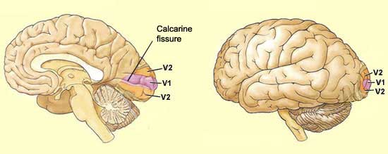
Primary brain areas studied
Drug use, specifically nicotine, effects three main portions of the brain. Those associated with:
• Reward – caudate nucleus
• Motivation – orbitofrontal cortex
• Visuo-spatial attention – parietal lobe, parahippocampal, & fusiform gyrus.
During a period of nicotine abstinence, mesolimbic and meso cortical (mesocorticolimbic brain system) dopamine circuits are stimulated.
The experiment
10 individuals (six men, four women) were recruited through public poster advertisements.
• 18-50 years of age
• Smoker for at least 2 years
• Smoke at least 10 cigarettes a day
• Average subject: 13.7 cigarettes per day, 8.1 years.
Passive treatment: participants were instructed to sit in the laboratory for 10 minutes. They were not allowed any kind of distraction (reading material, cell phones, internet, etc.) This amount of time was chosen because previous studies revealed this amount of time triggered nicotine cravings in similarly qualified test subjects.
Exercise treatment: participants were instructed on how to use a cycle ergometer. They then spent 2 minutes warming up on the bike and 10 minutes at a subjective
Rating of Perceived Exertion amounting to a light-to-moderately hard workout for each person. Their heart rate was monitored throughout.
After each treatment, the individuals were then led to the functional Magnetic Resonance Imaging (fMRI) scanner.
Before, during, and after each treatment and MRI scanning session, each person was asked to rate their desire to smoke from 1(none) to 7(strong desire).
MRI images

For approximately 15 minutes, those being tested viewed 60 images: 30 smoking-related images (hands holding cigarettes, lit cigarettes, etc.) and 30 neutral images (hands holding, people not smoking, etc.). Images were matched for color contrast and size. In between each image was a white screen with a black fixation cross that allowed for each subject to remain focused. These images lasted randomly for 8, 10, and 12 seconds.
Results

MRI scans revealed that the reward-processing brain areas were indeed stimulated in the control subjects (those not exercising). Stimulated precentral gyrus, parahippocampal gyrus (learning and memory) and caudate areas imply that these individuals experienced an enhanced perceived feeling of pleasure from the images being presented.
Post-exercise, however, revealed significant activations in Broadmanns areas 8-10, the rostral-medial frontal region and posterior cingulated cluster. These results from MRI scanning imply that the amount of exercise performed by each person stimulated the brain in such a way that temporarily inhibited “certain brain regions that were not directly essential to performing and maintaining the exercise, or physiological homeostasis.” Post-exercise, individuals found stimuli less salient. As a result, the images were less likely to elicit cravings.
